 +91 63713 07504
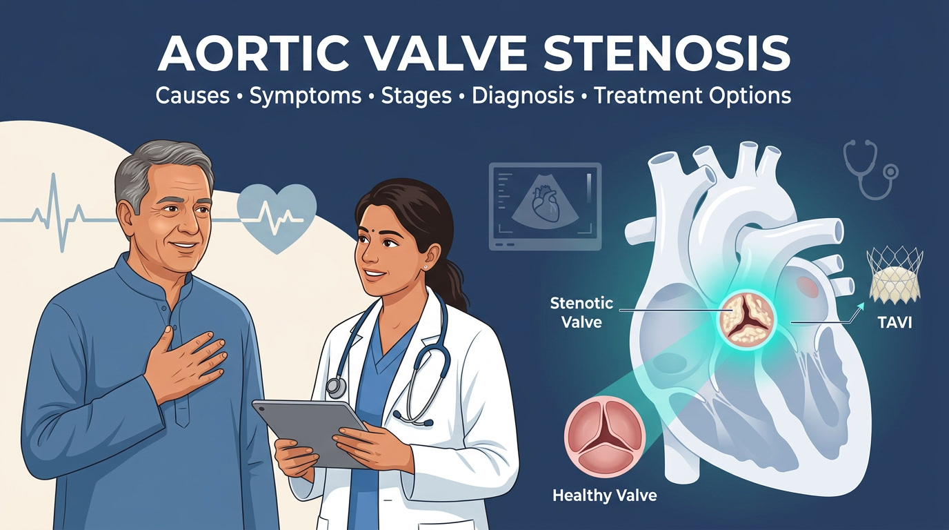
A narrowed aortic valve is one of the few heart conditions that can progress for years without producing a single symptom, and then, once it finally announces itself, change a person’s health trajectory within a matter of months. That gap between how quietly it begins and how seriously it can end is exactly why this condition deserves a far more thorough explanation than most patients ever receive during a ten-minute consultation.
Understanding what the valve actually does, why it narrows in the first place, how doctors measure that narrowing with precision, and what the evidence genuinely says about untreated disease gives a patient or a worried family member something a rushed appointment rarely can: the ability to ask the right questions and recognise when a decision genuinely cannot wait.
This guide is written to cover that entire arc in one place, from the underlying biology through every treatment option currently available, so that nothing essential about this condition is left unexplained.
Index: jump to any section
- Can aortic stenosis be reversed with diet, exercise, or medication?
- How long can someone live with severe aortic stenosis without treatment?
- Is a heart murmur always a sign of aortic stenosis?
- What is the difference between aortic sclerosis and aortic stenosis?
- Can young people get aortic stenosis?
- Does mild or moderate aortic stenosis need treatment right away?
- Is TAVI or open-heart surgery better for treating aortic stenosis?
What Is Aortic Valve Stenosis?
The heart relies on four valves to keep blood moving in a single direction with every beat, and the aortic valve is the last of these, positioned at the exit point where blood leaves the heart to supply the rest of the body. In its healthy state, this valve consists of three thin, flexible flaps, referred to as a tricuspid aortic valve, which open completely with each contraction and close fully afterward to prevent blood from flowing back into the heart.
Aortic stenosis develops when this opening becomes narrower than it should be, and the consequence of that narrowing is more significant than it might first appear. Because the passage through which blood must travel has become smaller, the heart’s main pumping chamber, known as the left ventricle, is forced to generate considerably more pressure with every single heartbeat in order to push the same volume of blood through.
Sustained over months and years, this additional workload causes the heart muscle itself to thicken, and the narrowing progressively reduces or blocks the flow of blood leaving the heart. This is a distinct condition from aortic regurgitation, in which the valve fails to close properly and blood is allowed to leak backward rather than being obstructed on its way forward.
What Causes Aortic Stenosis
Aortic stenosis is not a single disease with one origin story. Three distinct underlying mechanisms account for the overwhelming majority of cases, and identifying which mechanism applies to a given patient has real consequences, since it influences both the age at which the disease typically becomes serious and the direction the treatment conversation is likely to take.
Calcific Degeneration: the Most Common Cause Worldwide
In older adults, the leaflets of the valve gradually accumulate calcium deposits over the course of decades, a process that shares some similarities with how arteries develop plaque over time. This form of the disease is now recognised as the second-most frequent cardiovascular condition in developed countries after coronary artery disease and hypertension, and it typically becomes clinically significant from the sixth decade of life onward.
Bicuspid Aortic Valve: the Leading Cause in Younger Patients
A meaningful proportion of people are born with a valve that has only two flaps rather than three, a congenital variation known as a bicuspid aortic valve. Because a two-flap valve endures greater mechanical stress with every heartbeat across a lifetime, it tends to calcify and narrow considerably earlier than a normal valve does.
Patients with this congenital form typically require valve replacement in their forties, roughly two decades earlier than patients whose disease follows the standard calcific pattern, and although bicuspid valves are present in only around 1 percent of children, they account for nearly half of all valves ultimately removed for calcific stenosis in adult life.
Rheumatic Heart Disease: Still a Major Cause in India
This is the point at which the clinical picture diverges most sharply from what most Western medical literature describes, and it is particularly relevant for patients being evaluated in India. An untreated streptococcal throat infection during childhood can trigger rheumatic fever, an inflammatory response that gradually scars and thickens the heart valves over the years that follow.
Rheumatic aortic stenosis remains most prevalent in Asia, and India reports the highest burden of this form within the region. At a national level, the country carries an estimated 1.5 million cases of rheumatic heart disease, concentrated disproportionately in rural areas with limited access to timely medical care, and it tends to affect people during their most productive working years rather than in old age.
This is precisely why aortic stenosis encountered in Indian clinical practice can, and often does, appear decades earlier than the age at which Western guidelines assume it typically develops.
Other, Less Common Causes
Several less frequent causes also contribute to the overall picture, including congenital valves with only a single flap, certain genetic and metabolic conditions that affect calcium regulation in the body, chronic kidney disease, and prior radiation therapy directed at the chest.
Together, these account for only a small minority of cases, but they remain relevant considerations when a patient’s presentation does not fit the typical calcific or bicuspid pattern.
Who Is at Risk
Established risk factors for the calcific form of the disease include smoking, high blood pressure, elevated cholesterol, diabetes, and being male, all of which have been consistently identified across population studies. Age remains the single strongest driver within this category, with prevalence rising from roughly 2 to 3 percent among people over 65 to approximately 7 percent among those over 80.
For the rheumatic form of the disease, the strongest predictors identified in Indian research are low socioeconomic status and restricted access to antibiotic treatment for childhood throat infections, and the majority of affected patients in these studies fall between 15 and 44 years of age, a striking contrast to the elderly profile typically associated with valve disease in wealthier countries.
How Common Is Aortic Stenosis, Really
Exact figures vary depending on how a given study defines and measures the condition, but the underlying trend is remarkably consistent across every population studied: this disease is becoming more common as the world’s population ages. A large-scale Global Burden of Disease analysis spanning 204 countries and territories found that the overall burden of calcific aortic valve disease has climbed steadily over three decades, driven primarily by an aging population, with men consistently developing the disease at an earlier age than women.
Given continued gains in life expectancy, overall prevalence is projected to roughly double by the year 2050, and separate modelling suggests the number of patients over 70 living with calcific aortic stenosis will increase two to threefold across developed countries over the next fifty years.
How Aortic Stenosis Progresses Over Time
Aortic stenosis is, by its nature, a one-directional process. No currently available medication reverses valve calcification or reverses scarring once either process has begun. What clinicians can measure with reasonable precision, however, is the pace at which the disease tends to advance.
Once a patient’s stenosis reaches the moderate stage, research shows the valve opening typically narrows by roughly 0.1 square centimetres per year, accompanied by an average annual rise in pressure gradient of about 7 mmHg, and this pace of progression tends to accelerate in patients whose disease stems from a bicuspid valve or who already show heavier calcium buildup at the time of diagnosis.
This is precisely why a diagnosis of aortic stenosis is never treated as a single, static finding to be noted and set aside. Instead, it is followed through periodic echocardiograms over time, because a case classified as mild today can realistically progress to severe within a small number of years, particularly once moderate disease has already been established.
Symptoms: What to Actually Watch For
Perhaps the most consequential feature of this condition is how easily it can remain unnoticed for years at a time. Mild and even moderate stenosis frequently produce no perceptible symptoms whatsoever, largely because the heart muscle gradually thickens and compensates for the added workload without the patient noticing any change in daily function.
As a result, many people first discover they have the condition when a physician happens to hear an unfamiliar murmur during a routine physical examination, often long before any symptom would have prompted them to seek care on their own.
The Subtle, Early Signs
Before the more dramatic symptoms of advanced disease appear, patients often notice a handful of subtler changes that are easy to dismiss:
- A level of breathlessness during everyday activity that would not previously have caused any difficulty
- A degree of fatigue during ordinary tasks that feels disproportionate to the effort involved
- A general sense of gradually slowing down, which is frequently, and mistakenly, attributed entirely to normal aging
That last point deserves particular emphasis, since it is the one most likely to delay a diagnosis. As one Mayo Clinic cardiologist has observed, many older adults simply assume that reduced stamina reflects the natural course of getting older, when the more important question is whether that change actually represents ordinary deconditioning or an underlying, treatable cause that deserves proper evaluation.
The Classic Warning Triad
Once the valve narrows to a more advanced stage, three particular symptoms take on special clinical significance, since their appearance marks a genuine turning point in the disease’s trajectory, as explained in detail in the following section.
- Angina, meaning chest pain or a sensation of tightness, typically brought on by physical exertion
- Syncope, meaning fainting or a near-fainting sensation, particularly during activity
- Dyspnea, meaning breathlessness that progressively worsens toward the signs of heart failure, which can include swelling in the legs and difficulty breathing while lying flat
Less Obvious but Important Symptoms
A number of additional symptoms deserve attention even though they are less widely recognised as being connected to valve disease. Heart palpitations, dizziness brought on by exertion, and, in a subset of patients, chronic gastrointestinal bleeding can all occur as the condition progresses.
This bleeding tendency, occasionally referred to as Heyde’s syndrome, arises because the mechanical stress placed on blood as it forces its way through a narrowed valve gradually breaks down a clotting protein called von Willebrand factor, and it can present clinically as unexplained anemia, dark or tarry stools, or visible blood in the stool or vomit.
What Happens If Aortic Stenosis Is Left Untreated
This section tends to reshape how most people think about the condition once they read it, because the underlying research has remained remarkably consistent for more than five decades.
The Classic Findings
Researchers studying this disease as far back as the 1960s discovered that patients without symptoms survived at essentially the same rate as the general population while the underlying narrowing continued to progress unnoticed in the background.
Once genuine symptoms finally appeared, however, survival dropped sharply and without warning, a pattern the original investigators famously described as patients “falling off a cliff.”
That landmark study reported that average survival following the onset of symptoms was five years for patients who developed angina, three years for those who experienced fainting, and only two years for those whose first symptom was breathlessness.
A separate, large-scale European study conducted years later arrived at closely matching figures, finding that mean survival was 45 months after the appearance of angina, 27 months after fainting, and just 11 months after the first episode of heart failure.
For Patients Who Refuse or Cannot Have Surgery
The outlook for patients who decline recommended surgical treatment altogether is even more sobering. Among patients with severe stenosis who refused any form of surgery, mean survival was approximately 23 months, with only an 18 percent probability of surviving to the five-year mark, and every single patient in this group had died within twelve years of the initial observation period.
Why Doctors Watch "Asymptomatic" Severe Cases So Closely
Among patients who appear to have no symptoms despite already having severe stenosis, new symptoms have been documented in as many as 38 percent of cases within two years and 79 percent within three years. This is precisely why the phrase “no symptoms yet” is rarely treated by cardiologists as a reassuring long-term description of severe aortic stenosis. In practice, it functions far more like a waiting room than a diagnosis of safety.
Diagnosis: How Aortic Stenosis Is Actually Confirmed
Physical Examination
A heart murmur, described as a distinctive whooshing sound heard between heartbeats, is very often the first clue that leads to a diagnosis, and it can appear years before any symptom becomes noticeable to the patient. Because the earlier stages of this condition are genuinely subtle, most people with mild disease are identified during a routine checkup rather than because they sought care for feeling unwell.
Echocardiography: the Gold Standard
An echocardiogram serves as the standard initial test used to confirm and grade the severity of valve disease. This imaging study measures three interrelated values, and understanding what these numbers mean is genuinely useful for any patient trying to interpret their own report accurately.
| Severity | Aortic Valve Area (AVA) | Peak Jet Velocity | Mean Pressure Gradient |
|---|---|---|---|
| Mild | Greater than 1.5 cm² | 2.6 to 2.9 m/s | Less than 20 mmHg |
| Moderate | 1.0 to 1.5 cm² | 3.0 to 3.9 m/s | 20 to 40 mmHg |
| Severe | 1.0 cm² or less | 4.0 m/s or greater | 40 mmHg or greater |
These three measurements are considered together, and it is this combination that allows clinicians to classify the disease as mild, moderate, or severe with reasonable confidence.
When the Numbers Don't Agree With Each Other
There is an important nuance here that is rarely explained clearly to patients. In a meaningful proportion of cases, these three values fail to align neatly with one another.
Research indicates that roughly one-third of patients show some degree of mismatch between their valve area and pressure gradient, and the most common pattern involves a valve area small enough to suggest severe disease paired with a gradient low enough to suggest only moderate disease.
This mismatch typically arises because the heart is not pushing a sufficient volume of blood through the valve with each beat to generate a high gradient, even though the physical opening itself remains genuinely narrow.
When this kind of discordance is identified, physicians frequently turn to a CT scan that directly measures the amount of calcium buildup on the valve, since specific calcium score thresholds can help confirm whether the underlying stenosis is genuinely severe despite a reassuring gradient reading.
This distinction carries practical importance in everyday clinical care, since research has shown that women are disproportionately affected by this type of mismatch and are considerably more likely than men to have their true disease severity underestimated when gradient measurements are relied upon in isolation.
The CT Scan's Second Job: Procedure Planning
Beyond confirming disease severity, a detailed CT scan also plays an essential role once any form of valve intervention is being seriously considered, since it maps the precise dimensions of the valve and identifies the safest route for a catheter to travel through the blood vessels, a process explained in considerably more depth in our complete TAVR patient guide.
Other Supporting Tests
An electrocardiogram and a chest X-ray are typically performed alongside the echocardiogram to check for signs of heart strain or structural enlargement. In addition, blood tests measuring NT-proBNP, a hormone released by the heart when it is under mechanical stress, are being used with increasing frequency to help determine the appropriate timing of treatment in patients whose symptom status remains genuinely uncertain, a topic explored in further detail below.
The Official Staging System (ACC/AHA Stages A Through D)
Rather than relying solely on a simple mild, moderate, or severe label, current international guidelines apply a more detailed four-stage framework that accounts for valve anatomy, hemodynamic measurements, symptom status, and how the heart itself has responded over time.
| Stage | Meaning |
|---|---|
| A | At risk, with early valve changes but no true narrowing yet present |
| B | Progressive mild to moderate narrowing, without symptoms |
| C1 | Asymptomatic severe disease, with the heart still compensating normally |
| C2 | Asymptomatic severe disease, with early evidence of heart strain |
| D1 | Symptomatic severe disease, with a high pressure gradient |
| D2 | Symptomatic severe disease, with low flow, a low gradient, and reduced pumping function |
| D3 | Symptomatic severe disease, with a low gradient despite normal pumping function, corresponding to the mismatch pattern described above |
This more detailed breakdown of Stage D, defined by the interplay between gradient, blood flow, and ejection fraction, is what allows a heart team to match the urgency of treatment to a patient’s actual underlying physiology rather than relying on a single measurement in isolation.
The Role of Exercise Testing and Biomarkers
Patients labelled as asymptomatic despite having severe stenosis present a genuine diagnostic challenge, since some individuals have gradually and unconsciously reduced their level of activity over the years without ever noticing the change, effectively concealing symptoms that would otherwise have appeared.
This is precisely where a carefully supervised exercise stress test becomes clinically valuable, since it can reveal abnormalities that only emerge during physical exertion in patients who feel entirely well while at rest, and current guidelines recommend proceeding to valve replacement when such abnormalities are detected. It is worth noting that this same form of testing is considered unsafe, and is deliberately avoided, in patients who already experience symptoms while at rest.
Blood-based biomarkers add a further layer of useful information to this picture. Current guidelines already support earlier valve replacement in asymptomatic patients whose BNP levels are significantly elevated, on the reasoning that relieving the heart’s workload sooner may help prevent damage that would otherwise become permanent.
A 2025 analysis drawn from a major randomised trial found that higher levels of both NT-proBNP and troponin were associated with worse clinical outcomes overall, and notably, patients with normal troponin levels experienced a considerably greater relative benefit from early treatment than those whose troponin levels were already elevated.
Should Severe Aortic Stenosis Be Treated Before Symptoms Appear?
This question has become one of the most actively debated topics in modern cardiology, and the weight of evidence has shifted meaningfully in recent years. The traditional approach of waiting until symptoms actually appeared before intervening was grounded in that original 1960s observation that symptom-free patients had near-normal survival. A landmark clinical trial has since tested this long-standing assumption directly and rigorously.
In the EARLY TAVR trial, patients with asymptomatic severe aortic stenosis who underwent early transcatheter valve replacement experienced a lower combined rate of death, stroke, or unplanned cardiovascular hospitalisation compared with patients managed through standard surveillance and delayed treatment.
This finding represents a meaningful departure from the older, purely reactive model of care that dominated clinical practice for decades, and it helps explain why an increasing number of heart teams now raise the possibility of earlier intervention with carefully selected, symptom-free patients rather than defaulting to indefinite observation.
Every Treatment Option, Explained
Option 1: Watchful Waiting (Active Surveillance)
This approach is generally appropriate for patients in Stage A, Stage B, and carefully selected patients in Stage C1. It should not be mistaken for passive inaction, since it involves scheduled echocardiograms at defined intervals, often annually for moderate disease and roughly every six months for severe disease that remains without symptoms, alongside clear guidance on which symptoms warrant an immediate call to the doctor and active management of blood pressure and cholesterol levels throughout.
Option 2: Medical Therapy
It is worth stating this plainly, since it is a common source of confusion. No currently available medication reverses or even meaningfully slows the underlying calcification of the valve, and there are presently no drug therapies proven to alter the disease’s fundamental course.
Medications used in this setting are directed instead at managing blood pressure, cholesterol, and any heart failure symptoms that have already emerged, functioning as supportive care alongside ongoing monitoring rather than as a genuine substitute for intervention once the disease becomes both severe and symptomatic.
Option 3: Balloon Aortic Valvuloplasty (BAV)
This procedure uses a catheter-mounted balloon to stretch open the narrowed valve mechanically, without replacing it altogether. While it can provide meaningful relief in the short term, the improvement it produces is rarely durable. Large clinical studies have documented restenosis rates ranging from 40 to 80 percent within five to nine months of the procedure. For this reason, balloon valvuloplasty used as a standalone, permanent treatment is associated with meaningfully worse long-term outcomes than valve replacement, and in current practice it is used primarily as a temporary bridge before a patient proceeds to TAVI or surgery, or as an emergency measure in patients who are too unstable for immediate definitive treatment.
Option 4: Surgical Aortic Valve Replacement (SAVR)
This is open-heart surgery performed to remove the diseased valve entirely and replace it with either a mechanical or a biological prosthesis. It remains the preferred approach for younger patients at lower surgical risk, largely on the strength of its decades-long, well-documented track record for long-term durability.
The procedure requires opening the chest and carries a longer recovery period than catheter-based alternatives, but it remains the most thoroughly established valve treatment available anywhere in the world.
Option 5: Transcatheter Aortic Valve Implantation (TAVI/TAVR)
This is a minimally invasive, catheter-delivered valve replacement performed entirely without opening the chest. The full detail behind candidacy criteria, the step-by-step procedure itself, expected recovery timelines, the risk of requiring a pacemaker, and long-term durability data are all addressed comprehensively in our two dedicated guides on the subject.
Choosing Between These Options: The Heart Team Approach
No single test or measurement determines the appropriate treatment in isolation. Age, surgical risk scores, valve anatomy, kidney function, overall frailty, and the patient’s own personal priorities all factor into what is ultimately a shared decision made jointly between the patient and a multidisciplinary heart valve team, the same collaborative evaluation process described in considerable depth in our linked TAVI and TAVR guides above.
Living With Aortic Stenosis: Practical Guidance
For patients currently in the watching phase, meaning mild to moderate disease without symptoms, several practices are worth maintaining consistently. Every scheduled echocardiogram should be kept, even during periods when the patient feels entirely well, since the absence of symptoms is not a reliable indicator that the disease has stopped progressing.
Any new breathlessness, chest discomfort, or fainting should be reported to a physician immediately rather than held over for the next scheduled appointment. Blood pressure, cholesterol, and diabetes should all be actively managed, since each of these conditions has been shown to accelerate valve calcification. Finally, the presence of valve disease should be mentioned before any dental or surgical procedure, since valve tissue carries an elevated risk of infection during such interventions.
For patients who have already been diagnosed with severe stenosis, whether symptomatic or not, a slightly different set of priorities applies. Unsupervised, vigorous physical exertion should generally be avoided until a heart team has completed a full assessment, since severe stenosis can meaningfully limit the heart’s ability to increase its output on demand.
It is worth asking a physician directly whether an exercise stress test or BNP testing would be appropriate, particularly for anyone genuinely uncertain whether they are truly free of symptoms or simply accustomed to a gradually reduced level of activity. Perhaps most importantly, it is essential to understand that the absence of urgency is not the same as the absence of risk once a patient has reached Stage C2 or Stage D. These conversations with a cardiologist deserve to happen promptly, not to be deferred indefinitely out of a sense that nothing feels immediately wrong.
Aortic Stenosis in the Indian Context: What Makes It Different
Two features distinguish the picture in India from what most Western medical literature on this subject describes. The first is that India carries a genuine dual burden of disease, combining the classic, age-related calcific form seen throughout the developed world with a persistent rheumatic disease burden that continues to affect people well into their twenties, thirties, and forties.
On a global scale, rheumatic heart disease still accounts for more than 33 million cases and approximately 300,000 deaths annually, and it remains the most common cardiovascular disease requiring surgical treatment among young adults living in low and middle-income countries.
The second, closely related point is that age alone is a considerably less reliable indicator of the underlying cause of a given patient’s aortic stenosis in this setting than it might be elsewhere in the world.
A patient in their mid-thirties presenting with significant aortic stenosis represents a genuinely different clinical scenario in this region than the same finding would suggest under Western clinical assumptions, and this difference often shapes not only the presumed cause but also the eventual conversation around valve choice should replacement become necessary.
Frequently Asked Questions
Can aortic stenosis be reversed with diet, exercise, or medication?
No. Once the valve has narrowed as a result of calcification or scarring, no medication, supplement, or lifestyle change can reverse that underlying structural change. Lifestyle measures remain valuable for managing broader cardiovascular risk factors and overall heart health, but valve replacement remains the only intervention capable of restoring normal blood flow through a severely narrowed valve.
How long can someone live with severe aortic stenosis without treatment?
This depends heavily on whether symptoms have already appeared. Patients without symptoms often maintain survival close to that of the general population for a period of time, but once classic symptoms such as chest pain, fainting, or breathlessness develop, average survival without treatment has historically ranged from roughly one and a half to five years, depending on which symptom appeared first.
Is a heart murmur always a sign of aortic stenosis?
No. A great many murmurs are entirely harmless and require no treatment whatsoever. That said, any newly discovered murmur, particularly in someone over the age of 50, is worth investigating with an echocardiogram in order to rule out underlying valve disease.
What is the difference between aortic sclerosis and aortic stenosis?
Aortic sclerosis refers to early thickening or calcification of the valve that has not yet caused significant narrowing or any meaningful obstruction to blood flow, essentially representing the stage that precedes true stenosis. Not everyone diagnosed with sclerosis eventually develops stenosis, but regular monitoring is still recommended, since a meaningful proportion of patients do go on to progress.
Can young people get aortic stenosis?
Yes, most commonly as a result of a bicuspid aortic valve present from birth, or, particularly in the Indian context, as a result of rheumatic heart disease acquired during childhood or adolescence. Neither cause is confined to older adults.
Does mild or moderate aortic stenosis need treatment right away?
No. Mild and moderate stenosis are managed through ongoing monitoring rather than active intervention. Treatment is generally reserved for severe disease, typically once symptoms have appeared, or in select cases of severe disease without symptoms where testing indicates a genuinely elevated risk.
Is TAVI or open-heart surgery better for treating aortic stenosis?
There is no single, universal answer to this question, since the right choice depends on age, surgical risk, valve anatomy, and each patient’s own priorities regarding recovery time versus long-term valve durability. This exact comparison is addressed step by step in our dedicated TAVI guide and our TAVR patient guide.
The Bottom Line
Aortic stenosis is fundamentally a mechanical problem, and it requires a mechanical solution. It does not respond to medication in the way that high blood pressure or elevated cholesterol often can, which makes early and accurate diagnosis, combined with honest, ongoing tracking of symptoms, the single most important thing a patient genuinely has within their control.
More than fifty years of accumulated research point toward one remarkably consistent conclusion: this disease can remain silent for a considerable period, but once real symptoms finally emerge, the window for calm, unhurried decision-making narrows very quickly. Anyone who has been told they have a murmur, or who has noticed unexplained breathlessness, fatigue, or fainting, should not treat an echocardiogram as an overreaction. It remains the only reliable way to actually understand what is happening inside the heart.
